External Carotid Artery
General Structure of the Diagram
The diagram illustrates the External Carotid Artery and its main branches, distributed across specific anatomical regions of the neck and head. The arteries are represented in pink, while the skull and bony structures are shown in pastel tones to differentiate territories.
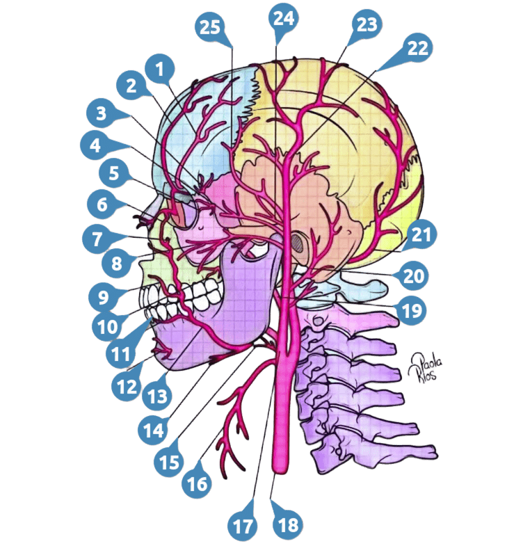
- Supraorbital Artery
- Supratrochlear Artery
- Zygomaticotemporal Artery
- Zygomaticoorbital Artery
- Zygomaticofacial Artery
- Angular Branch
- Infraorbital Artery
- Transverse Facial Artery
- Superior Labial Artery
- Buccal Artery
- Inferior Labial Artery
- Mental Branch
- Facial Artery
- Submental Artery
- Lingual Artery
- Superior Thyroid Artery
- External Carotid Artery
- Common Carotid Artery
- Maxillary Artery
- Posterior Auricular Artery
- Occipital Artery
- Superficial Temporal Artery
- Parietal Branch
- Middle Branch
- Frontal Branch
I. Superficial and Anterior Branches (Frontal and Orbital Region)
These arteries supply the forehead, eyelids, and zygomatic region:
- Supraorbital Artery
- Supratrochlear Artery
- Zygomaticotemporal Artery
- Zygomaticoorbital Artery
- Zygomaticofacial Artery
- Angular Branch (terminal branch of the facial artery)
Clinical Importance:
Essential in plastic surgery, facial trauma, and evaluation of orbital injuries.
II. Facial and Submental Branches (Middle and Lower Facial Region)
Located in the anterior part of the face, supplying the lips, cheeks, chin, and tongue:
- Infraorbital Artery
- Transverse Facial Artery
- Superior Labial Artery
- Buccal Artery
- Inferior Labial Artery
- Mental Branch (from the facial artery)
- Facial Artery
- Submental Artery
- Lingual Artery
- Superior Thyroid Artery
Clinical Importance:
Key in dental, maxillofacial procedures, and management of orofacial hemorrhages. The lingual artery is vital in tongue and floor of the mouth surgeries.
III. Main Trunk and Origin
Located in the lower cervical region, corresponding to the origin and trunk of the artery:
- External Carotid Artery
- Common Carotid Artery (common trunk giving rise to the internal and external carotid arteries)
Anatomical Note:
The bifurcation of the common carotid artery generally occurs at the level of the C4 vertebra.
IV. Posterior and Lateral Branches (Occipital and Auricular Region)
Supply the posterior part of the head, ear, and temporal region:
- Maxillary Artery (supplies deep facial structures, including masticatory muscles and teeth)
- Posterior Auricular Artery
- Occipital Artery
Clinical Importance:
The occipital artery is relevant in cranial surgery and the treatment of occipital headaches.
V. Terminal Branches of the Superficial Temporal Artery (Scalp)
Distributed in the superior and lateral region of the cranium:
- Superficial Temporal Artery
- Parietal Branch
- Middle Branch
- Frontal Branch
Clinical Importance:
These branches are fundamental in aesthetic, reconstructive surgery, and in the management of scalp wounds.